MRI Techniques
We are using and developing cutting-edge imaging techniques and post-processing algorithms to investigate brain volume, structure, connectivity, function, biochemistry, metabolism, and elastography.
MRI Modalities used at cBRAIN
Diffusion Weighted Imaging
[DESCRIPTION]
Principal Investigator:
Funding:
MR Elastography
Magnetic Resonance Elastography (MRE) is an advanced MR imaging technique that non-invasively characterizes and quantifies the mechanical properties of tissue. During MRE acquisition, shear waves are introduced into the brain using an external mechanical actuator attached to a passive pillow-driver underneath the participant’s head that vibrates at a continuous and known frequency. The resulting tissue deformations are encoded using specialized MR pulse sequences. The data is next run through an inversion algorithm to mathematically recover the mechanical properties of interest such as stiffness and damping ratio that are thought to reflect tissue composition and organization, respectively. Clinically, brain MRE is used to characterize brain tumours, and in research, has extensive relevance in the investigation of aging and neurodegeneration, traumatic brain injury, and other physiological and pathological processes. Our NEUROFIT study investigates brain health following repetitive head impacts using MRE (for more details see NEUROFIT).
People involved: Luisa Schuhmacher, Kate Rendall, Vishwa Singh, Alberto Villagran
External collaborators: Dr. Curtis Johnson and Olivia Bailey (Mechanical Neuroimaging Lab, University of Delaware)
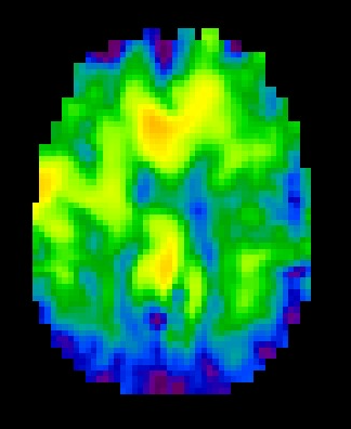
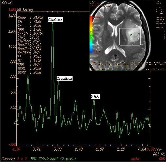
MR Spectroscopy
MRS is a non-invasive MRI technique that measures brain metabolites, providing insights into neuronal health, energy metabolism, and neuroinflammation. We use MRS to investigate metabolic alterations in traumatic brain injury (mTBI), as well as repetitive head impact (RHI), currently focusing on sex differences and hormonal influences.
By studying metabolites like N-Acetylaspartate, Creatine, Choline, and Lactate from specific brain regions, Anterior Cingulate Gyrus (ACG), Posterior Cingulate Gyrus (PCG), and Periventricular White Matter, we aim to uncover metabolic patterns in mTBI and RHI and the potential neuroprotective role of hormones, guiding targeted treatment strategies.